 0
|
0
|
 1
|
1
|
DEMINERALIZED BONE MATRIX (DBM)
biological scaffold | growth factor delivery | bone induction
__________________________________________________________
peer-reviewed sources only. all studies cited.
[1] Wildemann B et al. (2020) DBM in Bone Repair: History and Use
[2] Aghdasi B et al. (2013) DBM for Spinal Fusion: Evidence Review
[3] Brouwer RJ et al. (2017) DBM in Trauma and Orthopaedics: Systematic Review
[4] Eastlack RK et al. (2023) DBM and Fibers in Spinal Fusion
[5] Srivastava A et al. (2024) Spine Biologics: DBM Systematic Review 2014-2024
[6] Schwartz Z et al. (1998) Donor Age and Gender Effects on DBM Osteoinductivity
__________________________________________________________
CONTENTS
01 what is DBM
02 how it is processed
03 mechanism of action
04 growth factors: each one
05 clinical evidence
06 the variability problem
07 DBM vs other grafting options
08 commercial products
09 safety and risks
10 next generation DBM
11 verdict
__________________________________________________________
DEMINERALIZED BONE MATRIX
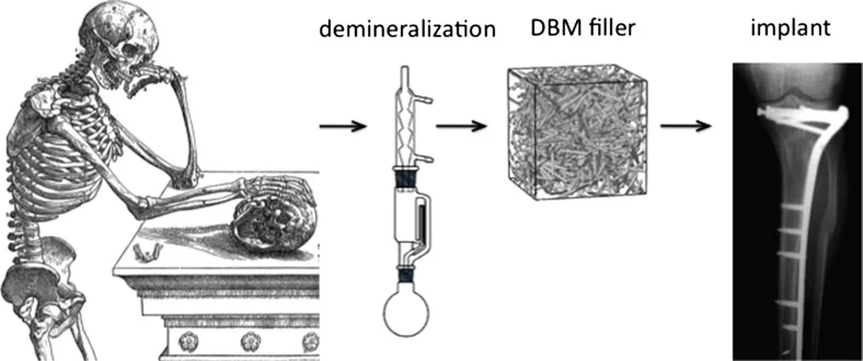
Allograft bone from a cadaveric donor with its mineral component
dissolved out by acid. What remains is the organic collagen matrix
plus the bioactive proteins that were embedded inside the bone.
Those proteins are growth factors. They recruit the patient's own
stem cells and direct them to differentiate into osteoblasts,
which then produce new bone at the implant site.
This is what separates DBM from synthetic substitutes like
hydroxyapatite or tricalcium phosphate. Synthetics are passive
scaffolds. DBM carries biological instructions.
__________________________________________________________
First described 1965, Marshall Urist (UCLA)
Key finding subcutaneous DBM in rabbits caused de novo bone formation
Clinical use since early 1970s
Global market share approximately 20% of all bone grafting procedures
US procedures/year approximately 108,000
__________________________________________________________
Available forms:
putty / paste / gel / flexible sheets / granules / powder
THE PROCESSING PIPELINE
Every step either preserves or degrades the biological activity
that makes DBM useful. This is also the source of the variability
problem covered in section 06.
__________________________________________________________
STEP 1 donor procurement
Cortical long bones (femur, tibia, fibula) from screened cadaveric donors.
Donors tested for HIV, hepatitis B/C, syphilis, HTLV.
Regulated as HCT/P under FDA 21 CFR Part 1271.
No pre-market clinical efficacy requirement for the final product.
STEP 2 cleaning and defatting
Soft tissue and marrow removed. Lipids extracted with organic solvents
or detergents. Strips most cellular material (HLA antigens), reducing
immunogenicity. Protocol varies by tissue bank. No industry standard.
STEP 3 demineralization
HCl at 0.5-0.6 N dissolves calcium hydroxyapatite.
The mineral normally traps growth factors inside the bone matrix.
Removing it exposes those factors for release in vivo.
Target residual calcium content: below 8% of dry weight.
Variables that determine product quality:
acid concentration / acid-to-bone ratio / temperature
agitation / contact time / particle size / residual calcium
STEP 4 sterilization
gamma irradiation effective sterility / denatures BMP and TGF-beta
electron beam (wet) reduces osteoinductivity by approximately 22%
ethylene oxide lower protein damage / sterility concerns at low dose
peracetic acid best balance of sterility and growth factor preservation
STEP 5 carrier addition
Raw DBM powder has no handling properties. Carriers added:
glycerol / sodium hyaluronate / gelatin / calcium sulfate
The carrier dilutes the active DBM content.
Carrier-to-DBM ratio is not required to be disclosed on the label.
TWO MECHANISMS
__________________________________________________________
OSTEOINDUCTION
The active biological mechanism. DBM releases growth factors into
the wound environment. Those factors recruit mesenchymal stem cells
from host periosteum, endosteum, and bone marrow. The same signals
drive MSC differentiation into osteoblasts. New bone forms in tissue
that was not previously bone. This is what synthetics cannot do.
__________________________________________________________
OSTEOCONDUCTION
The passive structural mechanism. The collagen matrix provides a
3D scaffold that bone-forming cells migrate along, attach to, and
use as a template for new bone matrix deposition.
DBM is weakly osteoconductive relative to HA scaffolds. Its collagen
matrix degrades quickly, which limits structural support but allows
complete replacement by host bone without permanent foreign material.
__________________________________________________________
SEQUENCE AFTER IMPLANTATION
1 DBM placed at defect site
2 BMP-2/7 and PDGF diffuse into surrounding tissue
3 MSCs recruited from host periosteum/endosteum/marrow
4 MSCs differentiate into osteoblasts under BMP/TGF-beta signals
5 osteoblasts migrate along collagen scaffold
6 osteoblasts synthesise osteoid (collagen I matrix)
7 FGF drives angiogenesis to sustain the process with blood supply
8 mineralisation of osteoid via calcium phosphate deposition
9 scaffold remodels and is replaced by host lamellar bone
GROWTH FACTOR PORTFOLIO
DBM releases a portfolio of proteins that work across different
phases of bone formation. Not a single molecule. A system.
__________________________________________________________
BMP-2 Bone Morphogenetic Protein 2
Primary driver. Recruits MSCs from surrounding tissue and drives
their commitment to the osteoblast lineage. Member of the TGF-beta
superfamily. Same protein used at supraphysiological doses in rhBMP-2
products (Infuse). In DBM it is present at natural physiological
concentrations. Content varies by donor age, processing, and product.
BMP-7 Bone Morphogenetic Protein 7 / OP-1
Synergises with BMP-2. Independently stimulates bone formation.
Also known as Osteogenic Protein-1. Amplifies the BMP-2 osteoinductive
signal and supports later-stage mineralisation.
TGF-beta1 Transforming Growth Factor beta-1
Confirmed present and functionally active in processed DBM via ELISA.
A 2021 study (OraGRAFT products) confirmed TGF-beta1 survives standard
demineralization, virus inactivation, and sterilization steps.
Drives osteoblast proliferation and matrix synthesis. Modulates immune
environment around the implant, reducing inflammatory scaffold degradation.
IGF-1 and IGF-2 Insulin-like Growth Factors
Promote osteoblast survival, proliferation, and bone matrix synthesis.
Synergistic with BMPs. IGF-1 has a well-characterised role in coupling
bone resorption to formation during normal remodelling cycles.
FGF Fibroblast Growth Factor
Drives angiogenesis at the graft site. New blood vessel formation
is required to sustain bone formation beyond the initial phase.
Without vascular supply, osteoblast activity stalls due to hypoxia
regardless of how many cells have been recruited.
PDGF Platelet-Derived Growth Factor
Recruits additional MSCs and stimulates early cellular proliferation.
Accelerates initial colonisation of the scaffold in the first days
after implantation.
Osteocalcin and Osteopontin
Non-collagenous bone matrix proteins. Regulate mineralisation of
newly formed osteoid and mediate osteoblast adhesion to the scaffold.
CLINICAL RESULTS BY APPLICATION
__________________________________________________________
CERVICAL SPINE FUSION (strongest evidence)
Kim et al 100% fusion rate, Grafton DBM alone in ACDF
Roh et al 97% fusion, Grafton DBM in LLIF
An et al allograft plus DBM equivalent to autograft in ACDF
prospective multicenter study
Gatam et al DBM plus HA: 76.5% fusion vs 77.8% autograft at 1 year
3 of 5 cervical spine studies reported 100% fusion.
All reported noninferiority vs autograft.
__________________________________________________________
LUMBAR SPINE FUSION (variable evidence)
Cammisa et al Grafton gel equivalent to autograft at 2 years
prospective controlled, side-by-side in same patient
Lee et al 73% fusion with Grafton alone in OLIF
Hyun et al RCT DBM gel vs DBM gel plus rhBMP-2
no significant difference in fusion at 11 months
Ouyang et al DBM plus concentrated bone marrow aspirate
80 patients, successful fusion, low complications
__________________________________________________________
ADULT SPINAL DEFORMITY
Bari et al (2022) demineralized cortical fibers associated with reduced
pseudarthrosis after pedicle subtraction osteotomy
Heegaard et al (2023) low pseudarthrosis in deformity surgery without 3-col osteotomy
__________________________________________________________
FRACTURE REPAIR
Geesink et al 5 of 6 critical fibular defects treated with DBM alone
first human confirmation of Urist's 1965 rabbit data
Standard threshold defects exceeding 2cm require intervention beyond host repair
__________________________________________________________
CRANIOFACIAL (2024)
Helsinki University Hospital, 138 patients, 2014-2022
DBX vs autogenous bone graft in Le Fort I osteotomies
orofacial clefts and craniofacial malformations
comparable complications and reoperation rates between groups
__________________________________________________________
SYSTEMATIC REVIEW 2024
Srivastava et al, 10 years of spinal fusion DBM data
conclusion: high fusion rates as graft extender or enhancer
consistent finding: DBM standalone less reliable than as component of strategy
THE VARIABILITY PROBLEM
10 to 15 percent of commercial DBM preparations have no meaningful
osteoinductive activity. There is no way to tell from the packaging.
__________________________________________________________
SOURCE 1 DONOR BIOLOGY
Younger donors yield DBM with higher BMP content and greater
osteoinductive capacity. Schwartz et al (1998): statistically significant
linear decline in osteoinductivity with increasing donor age in males.
In females the relationship is less consistent.
Optimal donor window: approximately 45-55 years (Alaribe et al 2016).
Neither age nor gender is screened for by most tissue banks.
SOURCE 2 DEMINERALIZATION VARIABLES
incomplete demineralization: growth factors remain trapped in residual mineral
over-demineralization: collagen architecture and protein content damaged
particle size: smaller particles produce more bone per unit area (Glowacki et al)
no industry-wide particle size standard exists
SOURCE 3 STERILIZATION DAMAGE
gamma irradiation denatures BMP and TGF-beta directly
e-beam in wet state reduces osteoinductivity by approximately 22% (Qiu et al)
sterilization protocol is not disclosed on the product label
SOURCE 4 CARRIER DILUTION
carrier-to-DBM ratio is not standardised and not required to be disclosed
a product that is 30% DBM and 70% glycerol delivers far less biological
signal than one that is 70% DBM. indistinguishable at point of purchase.
SOURCE 5 REGULATORY GAP
DBM regulated under FDA Section 361 as minimally manipulated human tissue
no pre-market efficacy data required
no standardised osteoinductivity testing requirement
no comparative performance requirement before reaching surgeons
17 different commercial DBM products on the US market.
Performance varies enormously. Most comparative data is manufacturer-funded.
BONE GRAFTING OPTIONS COMPARED
__________________________________________________________
AUTOGRAFT (iliac crest bone graft)
mechanism osteoinduction + osteoconduction + osteogenesis (live cells)
evidence gold standard. most reliable option.
advantage all three bone-forming mechanisms. no immune response. no disease risk
disadvantage donor site morbidity. limited volume. extra surgical time and blood loss
verdict S-TIER biologically. limited by harvest cost
FROZEN / FREEZE-DRIED ALLOGRAFT
mechanism osteoconduction only (live cells and proteins destroyed by processing)
evidence extensive use. well-characterised.
advantage abundant supply. structural support. predictable resorption
disadvantage no osteoinduction. entirely dependent on host biology
verdict A-TIER structural scaffold / B-TIER biological signal
DBM as graft extender
mechanism osteoinduction + osteoconduction (no live cells)
evidence high fusion rates as extender. variable as standalone.
advantage retains natural growth factor portfolio. no harvest morbidity
disadvantage batch variability. 10-15% batches not osteoinductive. regulatory gap
verdict A-TIER as extender / B-TIER as standalone
DBM + BONE MARROW ASPIRATE
mechanism DBM osteoinduction + BMA live MSCs + BMA growth factors
evidence 80-patient cohort: successful fusion, low complications (Ouyang et al).
advantage addresses the missing live cell component of DBM alone. synergistic
disadvantage BMA harvest adds a procedure step (less morbid than ICBG)
verdict A+ TIER practical clinical standard
rhBMP-2 (Infuse / Medtronic)
mechanism supraphysiological BMP-2 dose. powerful osteoinduction.
evidence strong RCT data in lumbar fusion and tibial nonunion.
advantage consistent potent effect. no donor variability
disadvantage ectopic bone. osteolysis. retrograde ejaculation in ALIF. $5-8k per kit. cancer signal
verdict S-TIER efficacy. risk profile requires careful indication selection
SYNTHETIC SUBSTITUTES (HA / TCP / CaSO4)
mechanism osteoconduction only. no biological signalling.
evidence adequate for small contained defects. inferior for large voids.
advantage unlimited supply. consistent. no disease risk. cheap
disadvantage zero osteoinduction. relies entirely on host osteoblasts arriving independently
verdict B-TIER scaffold. best for contained small defects with competent host biology
17 PRODUCTS ON THE US MARKET. KEY ONES:
__________________________________________________________
GRAFTON (Osteotech / Medtronic)
most studied DBM in the literature. the reference product.
Gel, putty, flex, crunch. Glycerol carrier. Most comparative data references
Grafton. 100% ACDF fusion rates in multiple series.
DBX (MTF / Synthes)
sodium hyaluronate carrier. widely used in craniofacial.
Putty and mix formulations. Used in the 2024 Helsinki craniofacial study.
ALLOMATRIX (Wright Medical)
DBM plus calcium sulfate. dual mechanism.
Adds osteoconductive component to osteoinductive DBM. Calcium sulfate
resorbs predictably, creating local porosity. Enhanced osteogenic
differentiation vs native cancellous bone alone in vitro.
ACCELL EVO3 (Integra)
high DBM content. minimised carrier dilution.
Competitive with Grafton in rat posterolateral fusion models.
OraGRAFT (LifeNet Health)
dental and oral surgery specific. best growth factor preservation data.
Confirmed to retain functionally active TGF-beta1 through all standard
processing steps via ELISA quantification (2021 study).
NOTE:
Do not apply Grafton efficacy data to a different product.
Each product has its own processing protocol and performance profile.
SAFETY PROFILE
__________________________________________________________
DISEASE TRANSMISSION
All donors screened per FDA and AATB standards for HIV, hepatitis
B/C, syphilis, HTLV. Demineralization has partial pathogen inactivation
effect. Additional sterilization reduces residual risk further.
Estimated HIV transmission risk from processed allograft:
approximately 1 in 1,670,000
No documented HIV transmission from commercially processed DBM
meeting current standards has been reported.
__________________________________________________________
IMMUNE RESPONSE
Demineralization removes most HLA antigen-bearing cellular components.
Residual type I collagen is poorly immunogenic (conserved across individuals).
No HLA matching required. No immunosuppression required.
Clinically significant rejection is rare and self-limiting when it occurs.
__________________________________________________________
PRION RISK (CJD)
Theoretical. Standard processing does not fully inactivate prions.
No documented case of CJD transmission from processed bone allograft.
Donor screening excludes individuals with known neurological disease.
__________________________________________________________
GRAFT FAILURE AND PSEUDARTHROSIS
The primary clinical risk is non-union, not toxicity.
A DBM batch with low osteoinductivity in a high-demand fusion
environment will result in fibrous non-union rather than bone bridge.
Failed fusion means revision surgery.
This is why the variability problem is a safety issue, not only efficacy.
ACTIVE RESEARCH DIRECTIONS
__________________________________________________________
MAGNETIC FIELD AUGMENTATION (2024)
Human DBM in critically sized rat cranial defects exposed to 1 Tesla
external magnetic field showed superior bone formation vs DBM alone.
Mechanism: piezoelectric properties of bone collagen generate electrical
signals under magnetic stimulation that activate osteoblast differentiation.
Journal of Materials Chemistry B (2024). Pre-clinical.
NANOPARTICLE SFRP-1 SILENCING PLUS DBM
Nanoparticle delivery of antisense oligonucleotides silences SFRP-1
gene locally. Combined with DBM scaffolding, activates Wnt/beta-catenin
at the graft site to amplify the response to DBM-released BMPs.
Connects WAY-316606 mechanism directly to DBM biology.
Springer Nature (2022). Pre-clinical.
STEM CELL LOADED DBM
DBM scaffolds pre-seeded with MSCs before implantation address the
absent osteogenesis component. Pig model (2014): DBM plus BMP-2 and
TGF-beta3 transfected BMSCs showed complete full-thickness cartilage repair.
Regulatory and manufacturing complexity limits near-term translation.
PROCESSING STANDARDISATION
Honsawek et al: osteoinductive potential increases with decreased
mineralisation (mouse model). Glowacki et al: particle size effect
confirmed (rat model). Both cited in ongoing regulatory push for
industry-wide processing standards. Progress is slow.
__________________________________________________________
WHAT DBM IS
Processed cadaveric bone retaining the growth factor content of native
bone after acid mineral extraction. Actively recruits and differentiates
bone-forming cells. Provides a collagen scaffold. Provides no live cells.
60 years of clinical history.
__________________________________________________________
STRENGTHS
graft extender alongside autograft in spinal fusion
bone void filling in fracture repair
cervical spine fusion (strongest standalone evidence)
no donor site morbidity vs autograft
combined with bone marrow aspirate addresses missing cell component
__________________________________________________________
LIMITATIONS
unreliable as standalone graft in high-demand biological environments
cannot replace autograft where live cells determine outcome
10-15% of batches have no meaningful osteoinductive activity
batch variability structurally unsolved due to regulatory gap
__________________________________________________________
A-TIER as graft extender
B-TIER as standalone graft
Proven biology. Real clinical utility. Meaningful limitations.
Best used as part of a strategy, not as the whole strategy.
__________________________________________________________
__________________________________________________________
educational reference. all information from peer-reviewed literature.
consult an orthopaedic surgeon for clinical decisions.
__________________________________________________________